


Coksarrosis- This is the osteoarthritis of the hip joint.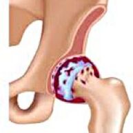 It develops gradually, for several years, prone to progression, it can be a smooth side and double.It is accompanied by pain and restriction of articulation movements.In the posterior stages, atrophy of the hip muscles and the shortening of the limb are observed.The diagnosis is established on the basis of clinical symptoms and radiography results.In the early stages of coxarthrosis, conservative treatment.With the destruction of the articulation, especially in young and medium age patients, surgery (endoprostics) is indicated.
General Information
Cooksarrosis (osteoarthrosis or deforming osteoarthritis of the hip joint) is a degenerative-dystophical disease.Usually, it develops at the age of 40 or more.It may be the result of various injuries and joint diseases.Sometimes it happens for no apparent reason.Cooksarrosis is characterized by a gradual progressive course.In the early stages, conservative treatment methods are used.In the posterior stages, the joint function can only be restored operational.
In orthopedics and traumatology, coxarthrosis is one of the most common osteoarthritis.The high frequency of its development is due to a significant load in the hip joint and the generalized prevalence of congenital pathology: joint dysplasia.Women suffer from cookardrosis a little more often than men.
The causes of cookardosis
Primary osteoarthritis (which arises for unknown reasons) and secondary (developed as a result of other diseases) of the hip joint.
Secondary cooksis can be the result of the following diseases:
- Hip joint dysplasia.
- Innate dislocation of the thigh.
- PERTES DISEASES.
- Aseptic necrosis of the thigh head.
- Infectious lesions and inflammatory processes (for example, hip joint arthritis).
- Lesions (traumatic dislocations, hip neck fractures, pelvic fractures).
CookStosis can be a smooth side or double.With primary coxarthrosis, there is often a concomitant lesion of the column (osteochondrosis) and knee joint (gonarrosis).
Risk factors
Among the factors that increase the probability of coxarcher development include:
- Constant load increase in the Board.Very often, observed in athletes in people with excess body weight.
- Circulatory disorders, hormonal changes, metabolic disorders.
- Column pathology (kyphosis, scoliosis) or stop (flat feet).
- Old age and senile.
- A sedentary lifestyle.
Cooksarrosis itself is not inherited.However, the child's child can inherit certain characteristics (metabolic disorders, structural characteristics of the skeleton and the weakness of the cartilage).Therefore, in the presence of blood relatives suffering from coxarchesis, the probability of the appearance of the disease increases slightly.
Patanatomy
The hip joint is formed by two bones: íleon and femoral.The head of the thigh is articulated with the acetabulum of the iliac bone, forming a peculiar "hinge".During the movements, the acetabulum remains motionless, and the femoral head moves in several directions, ensuring flexion, extension, abduction, traffic and rotating hips.
During the movements, the joint surfaces of the bones without obstacles slide with each other, thanks to the soft, elastic and durable hyalín cartilage that covers the cavity of the rotating cavity and the head of the thigh.In addition, the hyaline cartilage performs a shock absorption function and is involved in the redistribution of the load during movement and walking.
In the joint cavity there is a small amount of joint fluid, which plays the role of lubrication and provides nutrition of the hyaline cartilage.The joint is surrounded by a dense and strong capsule.Above the capsule there are large femoral and buttock muscles, which provide movements in the joint and, together with the hyalín cartilage, they are also shock absorbers that protect the joint of the lesions with failed movements.
With coxarthrosis, the joint fluid becomes thicker and more viscous.The surface of the hyaline cartilage dries, loses softness, cracks.Due to the roughness that has emerged, the cartilage during the movements is constantly injured by each other, which makes its thinning and aggravates the pathological changes in the joint.As coxarchesis progresses, bones begin to deform, "adapting" to greater pressure.Metabolism in the joint is deteriorating.In the posterior stages of coxarchesis, severe atrophy of the muscles of the sore limb is observed.
Coxarthrosis symptoms
The main symptoms of the disease include joint pain, inguinal region, thigh and knee joint.In addition, with cokesartrosis, rigidity of the movements and stiffness of the articulation, disturbance of the march, limp, atrophy of the hip muscles and the shortening of the limb on the side of the lesion.A characteristic feature of cooksarrosis is a restriction of kidnapping (for example, the patient is difficult when it comes to sitting in a chair).The presence of certain signs and its severity depends on the Coxarchesis stage.The first and most constant symptom is pain.
InFirst -degree coksarrosisPatients complain of periodic pain, which occurs after physical activity (prolonged race or walk).The pain is located in the joint, less frequently in the thigh or knee.After the break, it usually disappears.The first grade coxarche is not broken, the movements are preserved in its entirety, there is no muscular atrophy.
In the patient's X list suffering from first -degree coxarthrosis, minor changes are determined: moderate unequal narrowing of the joint gap, as well as bone growth around the outer or internal edge of the acetabulum in the absence of changes from the head and neck of the femur.
InCoksarrosis 2 degreesThe pain becomes more intense, often appears at rest, radiates in the thigh and groin.After significant physical activity, the patient with cookyrosis begins to limp.The volume of movements in the joint decreases: the abduction and internal rotation of the thigh is limited.
In X -ray images for second -degree coxarches, significant unequal narrowing of the joint gap is determined (more than half from normal height).The femoral head moves a little up, deforms and increases in size, and their contours become unequal.Bone growths with this degree of coxarcher appear not only in the internal, but also on the outer edge of the acetabulum and leave the cartilage.
InCoksarsis 3 degreesThe pain becomes constant, the concern of the patients not only during the day, but also at night.Walking is difficult, when it moves, a patient with cookyrosis is forced to use a cane.The volume of movements in the joint is very limited, the buttock muscles, the lower hips and legs are stunted.The weakness of the thigh extraction muscles becomes the cause of the deviation of the pelvis in the front plane and shortening the limb on the sore side.To compensate for shortening, a patient who suffers from cookyrosis, when walking, inclines the body to the sore direction.Because of this, the center of gravity changes, the load in the sore joint increases sharply.
In the radiographs for third grade coxarthrosis, acute narrowing of the articular gap, a pronounced expansion of the thigh head and multiple bone growth is detected.
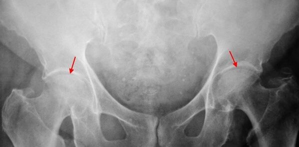
Diagnosis
The diagnosis of coxarthrosis is based on clinical signs and additional studies data, the main one of which is the radiography.In many cases, X -rays allow not only the degree of coxarcher, but also the cause of their occurrence.So, for example, an increase in the diffessional angle of the neck, the scenes and the flattening of the acetabulum indicate dysplasia, and the changes in the shape of the proximal part of the femur are indicated that cookardrosis is a consequence of the disease of pertes or the youth epiphysitic.In the radiographs of patients with coxarchesis, changes that indicate injuries can also be detected.
Like other methods of instrumental diagnosis of coxarchesis, TC and MRI can be used.Computed tomography allows you to study pathological changes in detail through bone structures in detail, and magnetic resonance images provide the opportunity to evaluate soft tissue disorders.
Differential diagnosis
In the first place, coxarthrosis should differentiate from gonarrosis (osteoarthrosis of the knee joint) and osteochondrosis of the spine.The atrophy of the muscles, which occurs in 2 and 3 stages of coxarcher, can cause pain in the knee joint, which are often expressed brighter than pain in the area of damage.Therefore, with the patient's complaints about the knee, a clinic (inspection, palpation, determination of the volume of movements) is the study of the hip joint, and if it is suspected of coxarcher, to direct the patient to the radiography.
Pain for root syndrome (compression of nerve roots) for osteochondrosis and some other diseases of the column can imitate pain with coxarthrosis.Unlike cookyrosis, when squeezing the roots, the pain occurs suddenly, after a failed movement, an acute turn, elevation of weights, etc., is located in the gluteus area and extends along the back of the thigh.A positive tension symptom is detected: severe pain when the patient tries to raise a straightened limb, lying on his back.At the same time, the patient freely carries his leg aside, while in patients with cookyrosis, the kidnapping is limited.It should be taken into account that osteochondrosis and cookyrosis can be observed at the same time, therefore, in all cases, an exhaustive examination of the patient is necessary.
In addition, cookesarchesis differs with trocanteritis (starting bursite) - aseptic inflammation in the area of junction of buttocks.Unlike coxarthrosis, the disease develops rapidly, within 1-2 weeks, usually after significant lesion or physical activity.The intensity of pain is greater than with cookyrosis.No limitations of the movements and the shortening of the limb are observed.
In some cases, with an atypical course of reactive disease or arthritis, symptoms can be observed that resemble coxarthrosis.Unlike coxarthrosis, with these diseases, the peak of pain falls at night.The pain syndrome is very intense, it can decrease when walking.The stiffness of the morning is characteristic, which occurs immediately after waking up and gradually disappears in a few hours.
COXARTROSIS TREATMENT
The treatment of pathology is dedicated to traumatologist orthopedists.The choice of treatment methods depends on the symptoms and the stage of the disease.In 1 and 2 stages of coxarthrosis, conservative therapy is carried out.During the period of exacerbation of coxarthrosis, injection blocks, non -steroidal anti -inflammatory drugs are used (pyroxes, indomethacin, diclofenaco, ibuprofen, etc.).It should be taken into account that the drugs of this group are not recommended for a long time, since they can have a negative effect on internal organs and suppress the hyaline cartilage capacity to restore.
To restore damaged cartilage for cookyrosis, funds from a group of condroprotectors (chondroitin sulfate, cartilage extract, etc.) are used.To improve blood circulation and eliminate the spasm of the small vessels, vasodilant drugs are prescribed (zinnararisine, nicotine acid, pentoxifilin, xantinol nicotinato).According to the indications, muscle relaxants (muscle relaxation drugs) are used.
With the stubborn pain syndrome, patients suffering from cookyrosis can prescribe intra -articular injections using hormonal drugs (hydrocortisone, triamcinolone, metrumor).Steroid treatment should be carried out with caution.In addition, with coxarthrosis, local products are used, heating ointments that do not have a pronounced therapeutic effect, however, in some cases relieve muscle spasm and reduce pain due to their "distraction" action.In addition, with coxarcherosis, physiotherapeutic procedures are prescribed (ultrasonic therapy, laser treatment, UHF, inducmia, magnetotherapy), massage, manual therapy and therapeutic gymnastics.
Diet for cookardrosis does not have an independent therapeutic effect and is used only as a means to reduce weight.The reduction of body weight allows you to reduce the load in the hip joints and, as a result, to facilitate the course of cooks.To reduce the load of the joint, the doctor, depending on the degree of coxarche, may recommend walking with a cane or crutches.
In the posterior stages (with third grade coxarthrosis), the only effective method of treatment is the operation: replace the destroyed joint with an endoprosthesis.Depending on the nature of the lesion, a single group can be used (replacing only the head of the thigh) or a group of two (replacing both the head of the thigh and the rotating cavity).
The endoprothetic operation for coxarthrosis is carried out in a planned manner, after a complete examination, under general anesthesia.In the postoperative period, antibiotic therapy is carried out.The seams are eliminated in 10-12 days, after which the patient is prescribed for an outpatient treatment.After endoprothetic, rehabilitation measures are necessarily maintained.
In 95% of cases, surgical intervention to replace the joint with coxarchesis guarantees a complete restoration of the function of the limbs.Patients can work, actively move and even practice sports.The average useful life of the prosthesis, subject to all recommendations, is 15-20 years.After this, a second operation is required to replace a worn endoprosthesis.