


Osteochondrosis is a disease of the spine, which is characterized by degenerative-dystrophic damage to the intervertebral discs, vertebral bodies and ligaments.
Osteochondrosis of the spine has a chronic progressive course.The disease does not manifest itself for a long time and symptoms appear only when complications arise.
According to statistics from the World Health Organization, 40% to 80% of the world's population suffers from osteochondrosis.
Among the patients, people over 30 years of age predominate.But lately there has been a trend towards rejuvenation of osteochondrosis.Osteochondrosis ranks first among spinal diseases in terms of disability among patients.
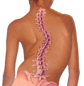
Brief anatomy of the spine.
The spine performs the main functions: the spinal cord canal, support and movement, and also connects the head, shoulders and pelvic girdle.
The structural unit of the spine is a vertebra.
The 24 vertebrae are connected to each other by intervertebral discs, which are the body's shock absorbers.
The spine is divided into five sections: cervical, thoracic, lumbar, sacral and coccyx.
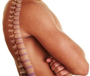
The normal shape of the spine is S-shaped.
This configuration of the organ allows the weight and body load to be evenly distributed.
Structural and functional elements of the spine.
A vertebra is a bone formation consisting of a body, an arch and processes.
The main load falls on the vertebral body, so this is its most massive part.
Important!The arches of adjacent vertebrae form the spinal canal, the spinal cord receptacle, blood vessels, spinal nerve roots, and adipose tissue.
LigamentsThe spine is represented by the posterior longitudinal ligament, which connects the vertebrae along the posterior surface, and the yellow ligament, the main purpose of which is to connect the arches of the vertebrae.
Vertebral processes.The vertebra has 7 processes extending from the arch: the spinous process, two transverse articular processes, two superior and two inferior.The ligaments and muscles of the spine are attached to the spinous processes.Other processes form the intervertebral joints of the spine.
intervertebral discIt is a disc-shaped plate consisting of a cartilaginous plate, an annulus fibrosus, and a nucleus pulposus.The intervertebral disc connects adjacent vertebrae, providing mobility and stability to the spine.
Intervertebral jointsFormed by the processes of two adjacent vertebrae.The main function of the intervertebral joints is to move the vertebrae relative to each other and provide flexibility to the spine.
intervertebral foraminaThey are found on the lateral sides of the spine and are formed by the articular processes, bodies and pedicles of the adjacent vertebrae.The roots of the spinal nerves exit through the intervertebral foramina and blood vessels enter.
spinal cord- This is a section of the central nervous system consisting of nerve fibers.The spinal cord has three membranes: soft, arachnoid and hard.The spinal dura mater consists of two sheets that connect and form the dural sac, filled with cerebrospinal fluid: cerebrospinal fluid.
spinal nerve roots- They are conductors of nerve impulses from the spinal cord to the internal organs and vice versa.Each spinal nerve root has autonomic, sensory and nerve fibers in its structure.
Paravertebral muscles- these are the muscles of the spine that support it and provide tilts and turns of the body.
The functional unit of the column isspinal movement segment, which consists of two adjacent vertebrae, an intervertebral disc, ligaments and muscles.
Pathogenesis (development mechanism) of spinal osteochondrosis.
In the process of development, osteochondrosis passes.four stages:
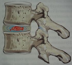
- First stage.Pathological changes do not extend beyond the boundaries of the intervertebral disc.The nucleus pulposus dries out, causing a decrease in the height of the intervertebral disc.The annulus fibrosus cannot withstand the load: it cracks and breaks.
- Second stage.Due to a decrease in the height of the intervertebral discs, sagging of the ligaments and muscles of the spine occurs, which leads to instability of the motion segment of the spine.The vertebrae can slide and move relative to each other.In this case, spondylolisthesis is formed.
- Third stage.The disease is progressing.Protrusion of the intervertebral discs and osteoarthritis of the intervertebral joints, as well as the uncovertebral joints, occur.
- Fourth stage.At this stage, adaptive reactions are activated in the form of bone growths of the vertebral bodies (osteophytes).Thus, the body tries to limit excessive mobility of the vertebrae.Osteophytes with their sharp edges damage the roots of the spinal nerves.Fibrous ankylosis of the intervertebral discs and joints forms and the spine becomes immobilized.The ankylosis stage is characterized by the disappearance of pain.
What leads to osteochondrosis?
Osteochondrosis of the backIt is a multifactorial disease in which it is impossible to identify a specific cause.
The basis of osteochondrosis is a violation of microcirculation and metabolism in the tissues of the spine, which can arise due to improper distribution of the load on the spine.
Factors contributing to the development of osteochondrosis include the following:
- incorrectly formed posture in childhood (scoliosis, kyphosis, kyphoscoliosis, slouching);
- weakness of the back muscles (incompetent muscular corset of the spine);
- staying in one position for a long time (working at a computer, working in an office, doing crafts);
- improper weight lifting;
- physical inactivity and sedentary lifestyle;
- metabolic pathology, especially lack of calcium, phosphorus, calcium, vitamins, magnesium, zinc;
- genetic predisposition to osteochondrosis;
- infectious diseases;
- frequent hypothermia of the body;
- chronic stress;
- hormonal imbalance;
- weightlifting;
- spinal injury;
- overweight and obesity.
Symptoms of osteochondrosis
Chronic osteochondrosis can manifest itself with various symptoms.It all depends on the stage of the disease, the level of damage to the spine and the presence of complications.
Clinically, the disease manifests itself when the degenerative-dystrophic process has already reached the posterior part of the annulus fibrosus and the posterior longitudinal ligament, then they become irritated, pinch the roots of the spinal nerves, and the conduction of nerve impulses through them is disturbed.
At the same time, compression of the spinal cord and blood vessels occurs, which is manifested by reflex and compression syndromes.
Important!Pain syndrome in osteochondrosis occurs due to pinching of the roots of the spinal nerves in the intervertebral foramina by osteophytes, spasmed muscles and displaced vertebrae.
Osteochondrosis with its symptoms often imitates acute coronary syndrome, pleurisy, acute pancreatitis, hepatic and renal colic, acute appendicitis and adnexitis.
Therefore, it is important to perform a thorough differential diagnosis of the disease to exclude life-threatening conditions.
Most commonSymptoms of osteochondrosis:
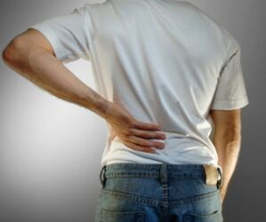
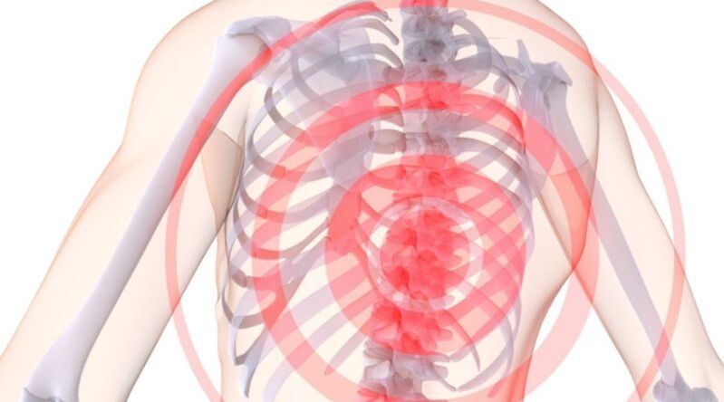
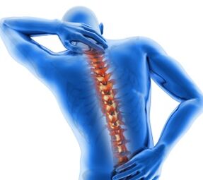
- pain in the neck, lower back, thoracic spine, which can be painful, throbbing or in the form of lumbago.The pain radiates to the head, upper and lower extremities, shoulder blades, heart and stomach.The pain syndrome increases after physical activity, sneezing, laughing, coughing or staying in the same position for a long time;
- sensory disturbancedifferent parts of the body at the level of innervation of the pinched nerve;
- spasmmuscles of the neck, back, upper and lower extremities;
- migraine type headaches;
- painsin the joints of the extremities;
- increased fatigueof physical and mental work;
- dizziness and loss of consciousnesswith a sharp turn of the head (vertebral artery syndrome);
- visual impairment(floaters before the eyes or colored spots);
- decreased hearing acuity, tinnitus;
- pain in the heart;
- painalong the intercostal spaces;
- decreased blood supplyupper and lower extremities, which is manifested by coldness of the skin;
- paresthesia– tingling, tingling and burning sensation in the spine;
- dry skin;
- sweating disorder;
- urinary disorder(dysuria, enuresis);
- decreased sexual desire, impotence.
Early diagnosis of osteochondrosis will greatly facilitate its treatment.
Methods for diagnosing osteochondrosis.
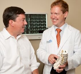
A neuropathologist diagnoses osteochondrosis.If necessary, patients can be referred for consultation to a cardiologist, gastroenterologist, orthopedist, surgeon and others.
During the interview, it is necessary to accurately determine the nature of the complaints, when they arose and what the patient associates them with.Be sure to check the patient's medical history, profession, and whether any close relatives have osteochondrosis.
Laboratory tests in this case are not informative.When conducting a biochemical blood test, you can pay attention to the level of calcium, phosphorus and other trace elements.
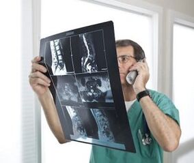
The main place in the diagnosis of osteochondrosis is occupied by instrumental methods, such as x-ray of the spine, computed tomography and magnetic resonance imaging.
Radiological examination of the spine is the simplest, most accessible and very informative method for diagnosing osteochondrosis.
Mandatory radiography is performed in direct and lateral projections of the desired part of the spine.Osteochondrosis is characterized by: decreased height of the intervertebral discs, presence of osteophytes, osteoporosis and spinal deformity.
myelography- This is a radiological examination of the spine with the introduction of a contrast agent into the spinal canal.This method is dangerous due to the occurrence of allergic reactions to the contrast.
Myelography allows us to study the internal structure of the spinal canal.The method is valuable in diagnosing Schmorl's hernias (intervertebral hernias).
Computed and nuclear magnetic tomography.– these are modern diagnostic methods that visualize the soft tissues and bones of the spine layer by layer.
These methods are expensive, so they are used in severe cases, especially for the differential diagnosis of osteochondrosis and diseases with similar symptoms.
Since osteochondrosis often disguises itself as diseases of the heart, lungs, pleura, stomach, intestines, kidneys and liver, it is necessary to carry out a differential diagnosis.
To do this, the patient may be prescribed an electrocardiogram, an ultrasound of the heart and internal organs, a blood test for troponins, an ultrasound of blood vessels, a chest X-ray, electroencephalography and others.
Treatment methods for osteochondrosis.
Treatment of osteochondrosis can beconservative and surgical.
Important!First of all, comprehensive conservative methods are used and surgical treatment is only resorted to in extreme cases.
Let's consider how osteochondrosis can be properly treated.kconservativeTreatment methods for osteochondrosis can be listed:
- drug therapy;
- physiotherapy;
- physiotherapeutic methods;
- manual therapy;
- massage;
- acupuncture.
Pharmacological treatmentOsteochondrosis is aimed at relieving pain, relaxing muscles, relieving swelling of nerves and muscles, improving blood flow and conduction of nerve impulses.For this, the following groups of drugs are used:
- non-steroidal anti-inflammatory drugs;
- chondroprotectors, which include components of cartilaginous tissue.These drugs protect the cartilage of the vertebrae and intervertebral discs from the negative effects of various factors;
- diureticswhich remove excess fluid from the body and relieve swelling of the roots of the spinal nerves and paravertebral muscles;
- muscle relaxantsrelax cramped muscles;
- drugs, improving metabolism and microcirculation in the spinal tissues (vitamins B1, B6, B12, C, A and E);
- calcium supplements;
- hormonal drugs, which are prescribed when non-steroidal anti-inflammatory drugs are ineffective.
therapeutic exercise– These are dosed physical activities that can be done both at home and at work for the treatment and prevention of osteochondrosis.
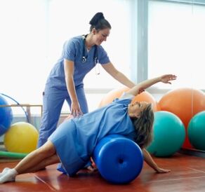
There are many sets of exercises for osteochondrosis.Prescribing exercise therapy and monitoring its implementation is carried out by a qualified specialist - a physiotherapist.
Thanks to correctly selected exercise therapy, it is possible to relieve pain, improve mobility and blood supply to the spine, and stop the progression of the disease.
Physiotherapeutic treatmentOsteochondrosis is carried out in special physiotherapy departments of hospitals, sanatoriums and dispensaries by a physiotherapist.
Physiotherapeutic methods include: electrophoresis, magnetotherapy, laser therapy, mud therapy, balneotherapy, ultraviolet exposure to the affected part of the spine, vibration treatment and others.
manual therapy– this is a dosed manual impact on the spine to restore its mobility, eliminate the displacement of the vertebrae and intervertebral discs.
Manual therapy should only be performed by a qualified chiropractor.
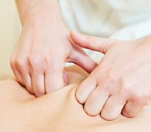
Massage and self-massagein case of osteochondrosis, it is performed to relieve muscle spasms, improve microcirculation in paravertebral tissues and increase spinal mobility.
acupunctureis a method of treating osteochondrosis in which fine needles are injected into active points.
Under the influence of needles in the body, the level of endogenous opiates and cortisol increases, which have anti-inflammatory and analgesic effects.
Prevention of osteochondrosis
To maintain your health and keep your spine mobile until old age, follow several principles for the prevention of osteochondrosis:
- watch your posture– always keep your back straight, do not slouch;
- choosecorrect postureto sleep;
- sit correctly at the table(relaxed shoulders, straight back, furniture should adapt to your height);
- during prolonged stay in one position (working in an office, at a computer, doing crafts), try every 1-1.5 hoursdo some physical exercise, back self-massage or just get up and walk;
- distribute the load correctlyin the spine when lifting and carrying various weights;
- wear orthopedic shoes;
- healthy sleepon a flat, hard to medium hard mattress.It is better to buy an orthopedic mattress and pillow.
Osteochondrosis of the spineIt is a chronic progressive disease that, unfortunately, cannot be cured.The effectiveness of the treatment depends directly on its punctuality.
Do not self-medicate so as not to worsen your condition.At the first signs of osteochondrosis, contact a neurologist.